How Is ABUS Different from Mammography?

Automated Breast Ultrasound (ABUS) and mammography are two distinct imaging modalities used for breast cancer screening and diagnosis. While both aim to detect abnormalities within the breast tissue, they differ significantly in their technology, methodology, and specific applications. Here’s an overview of how ABUS differs from mammography:
Technology and Methodology
Mammography:
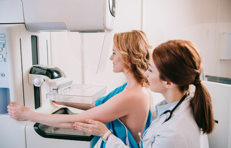
Mammography utilizes low-dose X-rays to create images of the breast. During the procedure, the breast is compressed between two plates to spread the tissue evenly, allowing for clearer images. Mammograms can show calcifications, masses, and other changes in the breast tissue that may indicate the presence of cancer. It is particularly effective in detecting early-stage cancers.
Automated Breast Ultrasound (ABUS):
ABUS uses high-frequency sound waves to create detailed images of the breast. Unlike handheld ultrasounds performed by a technician, ABUS is automated, providing a more consistent and comprehensive scan. During the procedure, the patient lies on their back, and a specialized transducer is moved across the breast, capturing multiple images that are then assembled into a 3D representation of the breast tissue.
Read also: Can Fruits Help improve Men’s Health?
Sensitivity and Specificity
Mammography:
Mammography is highly effective for most women, particularly those with fatty breast tissue. However, its sensitivity decreases in women with dense breast tissue, as dense tissue can mask tumours, making them harder to detect. This limitation is significant, given that dense breast tissue is relatively common.
ABUS:
ABUS is particularly beneficial for women with dense breast tissue. Dense tissue and tumours both appear white on mammograms, making it difficult to distinguish between the two. However, on an ABUS scan, dense tissue and abnormalities can be differentiated more clearly.
Applications
Mammography:
Mammography is the standard screening tool for breast cancer and is recommended annually or biennially for women starting at age 40-50, depending on individual risk factors and guidelines.
ABUS:
ABUS is typically used as an adjunct to mammography rather than a replacement. It is recommended for women with dense breasts, especially if they have additional risk factors for breast cancer. ABUS provides a more thorough examination in these cases, complementing the information obtained from mammograms.
Radiation Exposure
Mammography:
Mammography involves exposure to a small amount of ionizing radiation, which is a consideration for repeated screenings over many years. However, the benefits of early cancer detection generally outweigh the risks of radiation exposure.
ABUS:
ABUS does not use ionizing radiation, making it a safer option for repeated imaging, particularly for younger women or those who require more frequent monitoring.
Automated Breast Ultrasound and mammography are complementary tools in breast cancer detection. While mammography remains the gold standard for screening, ABUS offers significant advantages for women with dense breast tissue, improving the detection rates of abnormalities that mammography might miss. Together, these technologies enhance the overall effectiveness of breast cancer screening and diagnosis, providing a more comprehensive approach to breast health.


